

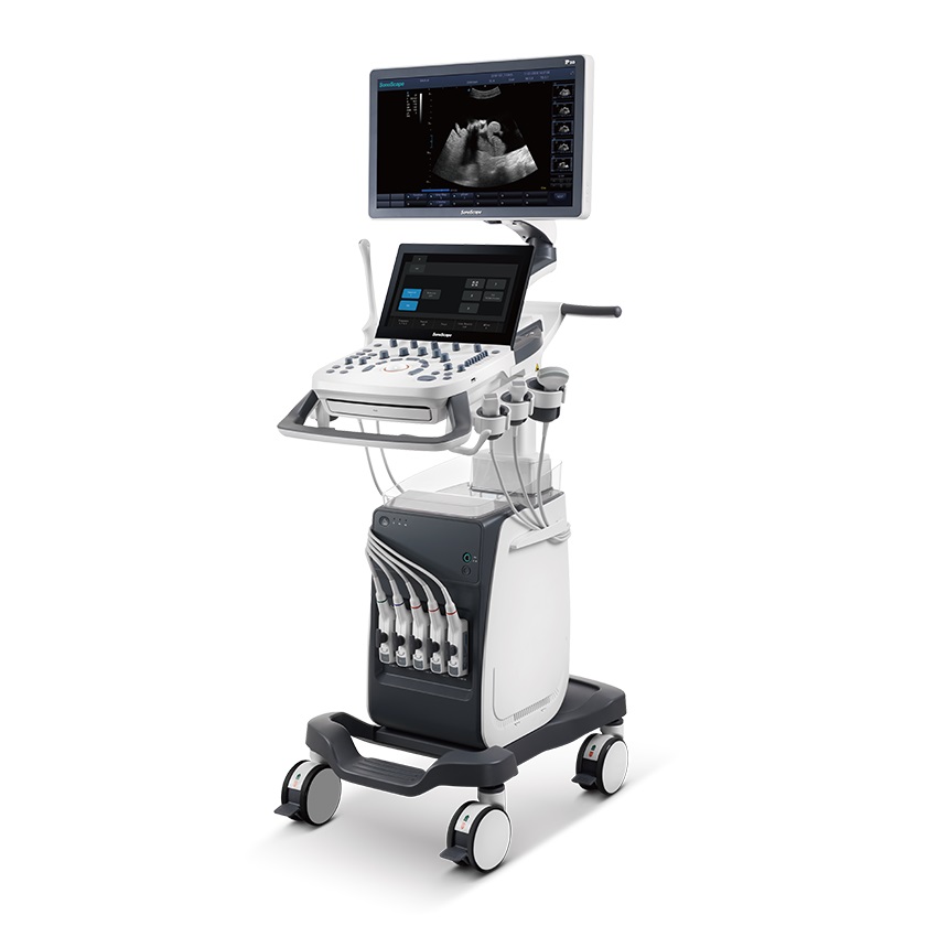

Incorporating innovative technologies, P20’s user-friendly designed with a simple operation panel, intuitive user interface and a variety of intelligent auxiliary scanning tools, will significantly improve your daily examination experience. Besides general imaging applications, P20 has entitled with diagnostic 4D technology which has an extraordinary performance in obstetrics and gynecology applications.
Specification
|
item
|
value
|
|
Model Number
|
P20
|
|
Power Source
|
Electric
|
|
Warranty
|
1 Year
|
|
After-sale Service
|
Online technical support
|
|
Material
|
Metal, Steel
|
|
Quality Certification
|
ce
|
|
Instrument classification
|
Class II
|
|
Type
|
Doppler Ultrasound Equipment
|
|
Transducer
|
Convex, Linear, Phased array, Volume 4D, TEE, Biplane Probe
|
|
Battery
|
Standard Battery
|
|
Application
|
Abdomen, Cephalic, OB/Gynecology, Cardiology, Transrectal, Peripheral vascular, Small parts, Musculoskeletal, Transvaginal
|
|
LCD monitor
|
21.5" High Resolution LED Color Monitor
|
|
Touch Screen
|
13.3 inch quick response
|
|
Languages
|
Chinese, English
|
|
Storage
|
500 GB Hard Disk
|
|
Imaging modes
|
B, THI/PHI, M, Anatomical M, CFM M, CFM, PDI/DPDI, PW, CW, T
|


Product Features
|
21.5 inch high definition LED monitor
|
|
13.3 inch quick response touch screen
|
|
Height-adjustable and horizontal-rotatable control panel
|
|
Abdominal solutions: C-xlasto, Vis-Needle
|
|
OB/GYN solutions: S-Live Silhouette, S-Depth, Skeleton
|
|
Auto Calculation and Auto Optimization Package: AVC Follicle, Auto Face, Auto NT, Auto EF, Auto IMT, Auto Color
|
|
Large capacity built-in battery
|
|
DICOM, Wi-fI, Bluetooth
|
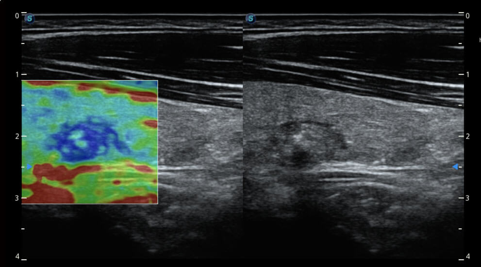
C-Xlasto Imaging
With C-xlasto Imaging, P20 enables comprehensive quantitative elastic analysis. Meanwhile, C-xlasto on P20 is supported by linear, convex and transvaginal probes, to ensure good reproducibility and highly consistent quantitative elastic results.
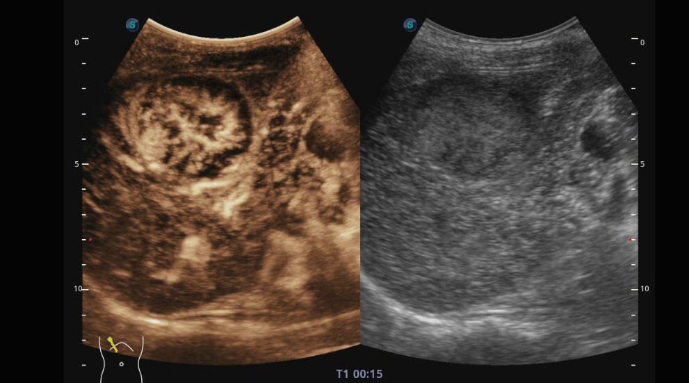
Contrast Imaging
Contrast Imaging with 8 TIC curves allows doctors to assess perfusion dynamics in a wide range of clinical settings, including both the location and evaluation of lesion parts.
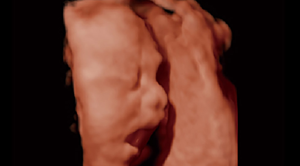
S-Live
S-Live allows for detailed visualization of subtle anatomical features, thereby enabling intuitive diagnosis with real-time 3D images and enriching patient communication.
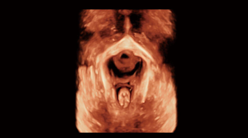
Pelvic Floor 4D
Transperineal 4D pelvic floor ultrasound can provide useful clinical values in assessing the vaginal delivery impact on the female anterior compartment, judging whether the pelvic organs are prolapsed or not and the extent, determining if the pelvic muscles were torn accurately.
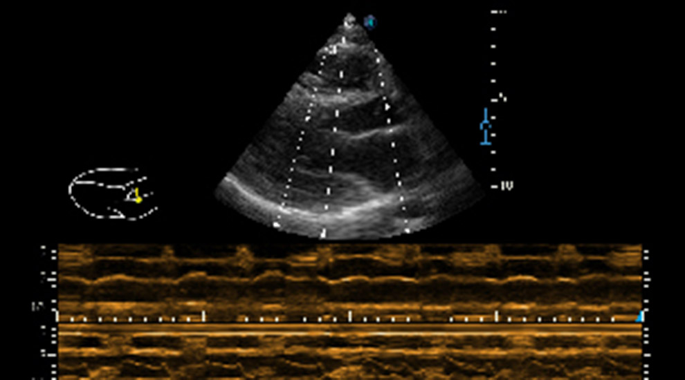
Anatomic M Mode
Anatomic M Mode helps you observe the myocardial motion at different phases by freely placing sample lines. It accurately measures the myocardial thickness and the heart size of even difficult patients and supports the myocardial function and LV wall-motion assessment.
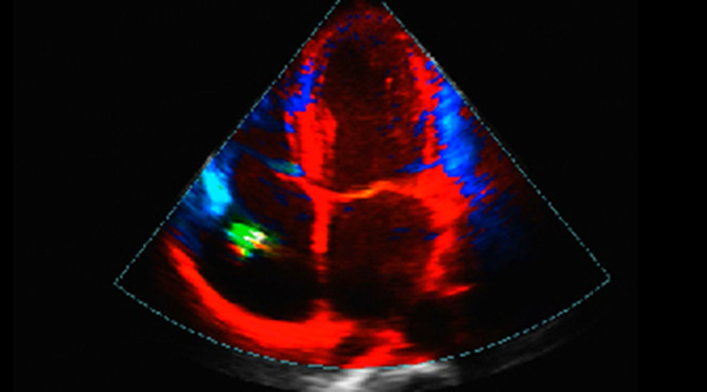
Tissue Doppler Imaging
P20 is endowed with Tissue Doppler Imaging which provides velocities and other clinical information on myocardial functions, facilitating clinical doctors with the ability to analyze and compare the motions of different parts of the patient's heart.


