


Specification
|
Basic Configuration
|
||
|
Advanced Imaging Technologies
|
Spatial Compound ImagingPulse Inversion Harmonic ImagingC-Xlasto elastographyReal-time 3D (4D)High Density Probe
|
|
|
Operation Modes
|
B, Dual B, Quad B, THI, Trapezoid Imaging, Real-time Panoramic Imaging(B mode and Color mode)
M, Color M, Anatomic M Stress Echo Color Doppler(the flow velocity can be calculated), Power Doppler Imaging, Directional PDI, TDI PW with HPRF, CW Dual-Live Duplex: B and Doppler/M, can be defined in new preset Triplex: B, Color Flow, and PW/CW Doppler,can be defined in new preset 3D Imaging 4D Imaging Contrast Imaging C-xlasto( elastography imaging) |
|
|
Applications
|
Abdomen OB/GYN
Cardiology Urology Small parts Vascular Orthopedic Pediatrics anesthesia MSK , etc. |
|
|
Monitor/Touch Screen
|
no less than 15 inch high resolution LED color monitor, the open angle is adjustable: 0°~50° / 13.3″ High Resolution Touch Screen
|
|
|
Transducers Sockets
|
At least 2 transdcuer connectors holders, which can support all transducers. 24 transducer choices, indcluding: linear, convex,
endocavity, phased array ,TEE, laparoscope and 4D transducers. |
|
|
Report Tables
|
Abdominal, OB/Gyn, Cardiology, Urology, Small Parts
|
|
|
Report Format
|
TXT, PDF
|
|
|
Report Templet
|
at least 6 images can be displayed in report
|
|
|
Doppler Cine Playback
|
Speed is adjustable; Sound can be played back.
|
|
|
User-define keys
|
can define the saving Image or saving Cine function
|
|
|
Scanning Methods
|
Electronic Convex Secter
Electronic Linear Secter Electronic Phased Array Sector |
|
|
ECG
|
support ECG function
|
|
|
Clipboard Function
|
Capture and review the archived images and cines
|
|
|
Image Optimization
|
Images can be optimized by one button in B/Color/PW mode
|
|
|
Screen Saver
|
0~99 minutes, adjustable
|
|
|
Built-in battary
|
can support 90 minutes continuously scanning
|
|
|
Weight
|
no more than 7.8Kg without built-in battery
|
|
|
Transducer Specification
|
||
|
Endocavity transducer
|
frequency range: 4~9MHz
scanning angle: 193° |
|
|
Temperature-detection Technology
|
the temperature of endocavity transducer can be displayed
|
|
|
Biplane
|
Biplane(Convex+Convex, Linear+Convex),dual active mode for Biplane Convex + Convex, biopsy grid for Biplane Linear+Convex
|
|
|
TEE
|
support TEE transducer both for adult and pediatrics
|
|
|
Phased Array
|
Low frequency for adult (1-5MHz)
High frequency for pediatrics (4-12MHz) Scan Ranges:≥90° |
|
|
Biopsy Guide
|
Required
|
|
|
Convex Transducer
|
frequency range:2~6MHZ scanning depth:3~240mm Scan Ranges:≥70°
|
|
|
Linear Transducer
|
elements:128/192/256
|
|
|
Optional Configuration
|
||
|
Configuration
|
Static 3D/4D
C-xlasto: Elastography Imaging ECG Module Hard Disk 1T |
|
|
Transducers
|
192 elements linear array L742(Vascular, Small parts, MSK etc.), 4-16MHz/ 38mm
192 elements linear array L743(Vascular, Small parts, MSK etc.), 4-16MHz/ 46mm 256 elements linear array L752(Vascular, Small parts, MSK etc.), 4-16MHz/ 52mm 128 elements linear array 10L1 (Vascular, Small Parts, MSK etc.), 4-16MHz/ 36mm 128 elements convex array 3C-A (Abdominal, Obstetrics, Gynecology), 1.0-7.0MHz/ R50mm 128 elements convex array C354 (Abdominal, Obstetrics, Gynecology), 2-6.8MHz/ R50mm 192 elements convex array C353 (Abdominal, Obstetrics, Gynecology), 2-6.8MHz/ R55mm 192 elements convex array C362 (Abdominal, Obstetrics, Gynecology), 2.4-5.5MHz/ R60mm 72 elements convex array C322(Abdominal Biopsy), 2-6.8 MHz/ R20mm 128 elements convex array C542 (Abdominal, Pediatrics), 3-15 MHz/ R40mm 128 elements micro-convex array C611(Cardiology, Pediatrics), 4-13 MHz/ R11mm 128 elements micro-convex array C613(Cardiology, Pediatrics), 4-13 MHz/ R14mm 80 elements phased array 4P-A (Cardiac, Transcranial), 1.0-5.4MHz adult 96 elements phased array 5P2 (Cardiac, Transcranial, Pediatric), 2-9MHz Pediatric 96 elements phased array 8P1 (Cardiac, Transcranial, Infant), 4-12MHz 128 elements endocavity 6V1 (Gynecology, Obstetrics, Urology), 3-15MHz/ R11mm 192 elements endocavity 6V3 (Gynecology, Obstetrics, Urology), 3-15MHz/ R10mm 128 elements endocavity 6V1A (Gynecology, Obstetrics, Urology), 3-15MHz/ R11mm 192 elements endocavity 6V7 (Gynecology, Obstetrics, Urology), 3-15MHz/ R10mm 96 elements linear array 10I2 (Intra-operative), 4-16 MHz/ 25mm 128 elements laparoscope linear array LAP7 (Intra-operative), 3-15MHz/ 40mm Volumetric convex array VC6-2 (Obstetrics, Abdominal, Gynecology), 2-6.8MHz/ R40mm PWD 2.0 (Cardiac, Transcranial), 2.0Mhz CWD 2.0 (Cardiac, Transcranial), 2.0MHz CWD 5.0 (Cardiac, Transcranial), 5.0MHz Transesophageal MPTEE (Cardiology), 4-13 MHz Transesophageal MPTEE Mini (Cardiology, Pediatric), 4-13 MHz 128 elements transrectal EC9-5 (Urology), 3-15 MHz/ R8mm 192/192 elements biplane BCL10-5 (Urology), Convex 3.9-11 MHz/ R10mm, Linear 6-15 MHz/ 60mm 128/128 elements biplane BCC9-5 (Urology), 3.9-11 MHz/ R10mm |
|
Product Features

*Spatial Compound Imaging
Spatial Compound Imaging utilizes several lines of sight for optimal contrast resolution, speckle reduction and border detection,
with which the S9 is ideal for superficial and abdominal imaging for better clarity and improved continuity of structures.
with which the S9 is ideal for superficial and abdominal imaging for better clarity and improved continuity of structures.
Pulse Inversion Harmonic Imaging
The harmonic signals are fully preserved without degradation of the acoustic information, which makes it possible for the S9 to
image high-level details and improve contrast resolution by reducing noise and clutter in the visualization of subtle lesions,
small parts, vascular and so on.
image high-level details and improve contrast resolution by reducing noise and clutter in the visualization of subtle lesions,
small parts, vascular and so on.
C-Xlasto elastography
SonoScape provides the S9 with a new method to support the physician in assessing tissue elasticity. The differences in tissue
responses are detected and visualized in real-time by the elastography algorithms through different graphical representations
which can be particularly helpful in analyzing breast, thyroid and musculoskeletal structures.
responses are detected and visualized in real-time by the elastography algorithms through different graphical representations
which can be particularly helpful in analyzing breast, thyroid and musculoskeletal structures.

Real-time 3D (4D)
With increased physical channels and a new platform, the S9 offers both high quality imaging and high frame rates to meet a new
standard of SonoScape’s S series. Thanks to high frame rate and advanced technologies, the 4D imaging of S9 delivers smooth
movement of the fetus and offers comprehensive 4D acquisition, data rendering, and post-processing functionality.
standard of SonoScape’s S series. Thanks to high frame rate and advanced technologies, the 4D imaging of S9 delivers smooth
movement of the fetus and offers comprehensive 4D acquisition, data rendering, and post-processing functionality.
High Density Probe
S9 is equipped with a high density phased array probe to meet the needs of both high frame rate and premium resolution in cardiac
imaging. Thanks to the high sensitivity of SonoScape’s color Doppler mapping, the S9 can provide an accurate cardiac diagnosis
beyond your imagination.
imaging. Thanks to the high sensitivity of SonoScape’s color Doppler mapping, the S9 can provide an accurate cardiac diagnosis
beyond your imagination.
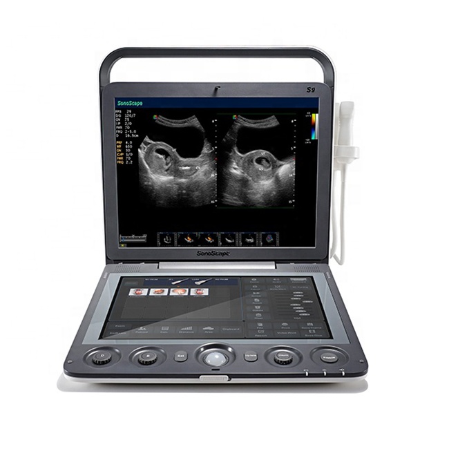
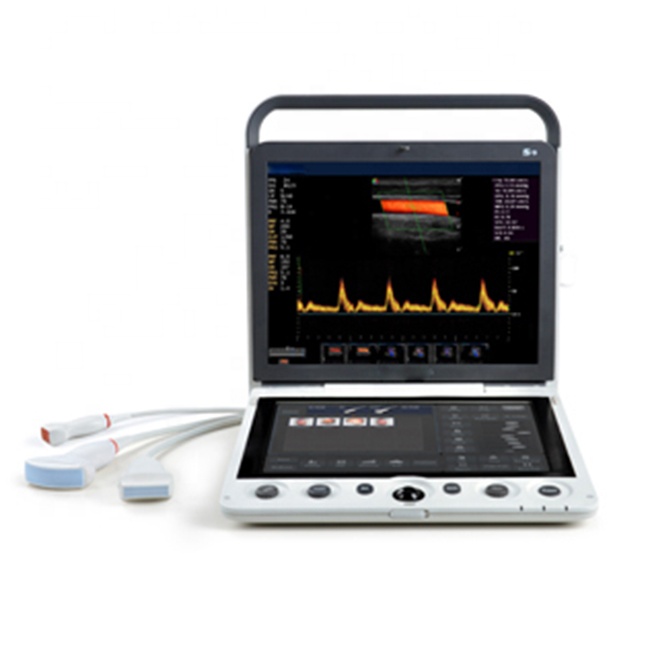
*15 inch high definition LED monitor
*13.3 inch touch screen
*Two transducer sockets
*Stylish trolley with adjustable height
*Removable built-in battery supports 90 minutes scanning per charge
*Full patient database and image management solutions: DICOM 3.0, AVI/JPG, USB 2.0, HDD, PDF report
*Premium application technology: u -Scan, Compound Imaging, Pulse Inversion Harmonic Imaging, TDI, Stress Echo, C-xlasto, and Contrast lmaging.
*A comprehensive selection of probes: Linear, Convex, Micro-convex, Endocavity, High-density phased array, Intraoperative, TEE,Bi-plane, Pencil, Volumetric, and Laparoscope probe
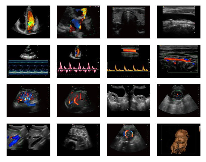
General Images
Leave Your Message:
Write your message here and send it to us.
-

Amain OEM/ODM waterproof Animal Pregnancy Port...
-

Amain OEM/ODM MPUL8-4E BW Transrectal Scanner P...
-

Amain professional Sow Pregnancy Tester Veterin...
-

Amain OEM/ODM MagiQ MPUC5-2ET endo-cavitary med...
-

Hd Digital wristband portable veterinary ultras...
-

Amain OEM/ODM MagiQ MPUEV9-4E Portable Veterina...


