

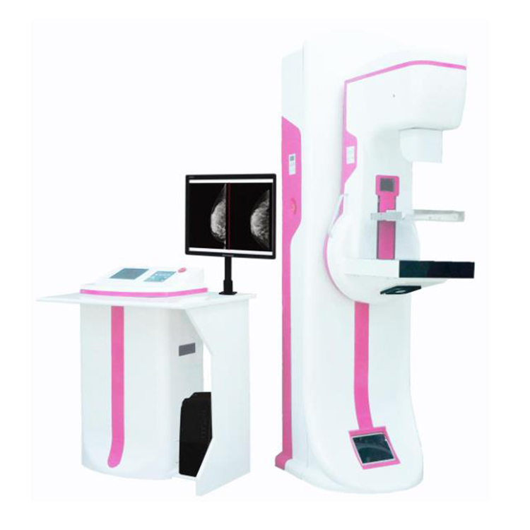
Amain Full Field Digital Mammography System Ce Approved Low-dose Original Factory Supply Digital X-ray System
Specification
|
Item
|
Parameter
|
Remark
|
|
X-ray Generator
|
Generator Type: High Frequency Inverter 80kHz
Input Power: Single phase 220VAC, 50/60Hz Radiographic Ratings: Large Focal Point 20-35kV/10-510mAs
Small Focal Point 20-35kV/10-100mAs
Power Rating: 6.2kVA |
Self-developed All-solid-state high frequency high voltage x-ray generator
|
|
X-ray Tube
|
Focal Spot Size: Dual Focus 0.1 / 0.3mm Target Material: Molybdenum (Mo)
Port Material: Beryllium (Be)
High-speed anode drive: 2800 /10000rpm Target angle: 10°/16°
Anode Heat Storage: 210kJ (300kHU)
Anode Cooling: Air cooling Filtration: Mo(0.03mm), Al(0.5mm)
|
Model:IAE C339V
|
|
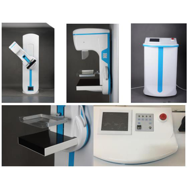
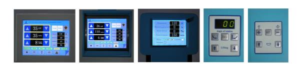
Radiographic Stand
|
C-ARM: Vertical Movement: 590mm
Center of electric rotating C-arm
Automatic return function by one key
Rotations Degree: +90°~-90° Automatically released after the exposure pressure settings display
Compression flexible, stepless speed.
Max. pressure: 200N
Max. travel: 150mm
SID: 650mm
|
Electric Isocentric rotating
|
|
Flat Panel Detector
|
Detector material: Amorphous silicon Effective coverage of detector: 24x30cm Pixel matrix: 3072×1944
Limit of spatial resolution: 6.0Lp/mm
DQE value: 70%
Dynamic range: 14bit digital output
Pixel size: 75μm
High voltage Synchronizer trigger: BNC Output: Camera Link or Ethernet
Working condition: 10℃-40℃
Storage environment: -10℃-50℃
|
|
|
Bucky housing and movement device
|
Size: 374*304*65mm
Stepless speed regulating range: 0~6cm/s
Movement range: 0.5~2cm
Grid Size: 24x30cm
Grid ratio: 5:1
Grid density: 30lp/cm
Focal distance: 650mm
|
|
|
Image acquisition workstation
|
CPU ≥ Intel Core Duo 2.60GHz
Hardware ≥ 250G high speed Hardware Memory ≥ 2G
Display card ≥ 512MB
High brightness high-contrast LCD,1280*1024 Pixel resolution
Network interface Work – list
DICOM 3.0 transmission
100/1000 Gigabit Ethernet
Software: Imaging software package DMOC V1.0
|
Configuration including Diagnose digital workstation
2M medical monitor for optional
|
|
Line Voltage
|
220V ac±10%@25A,Single phase
|
110V for optional
|
|
Packing Size
|
2160*690*1150mm
|
|
|
G.W
|
350kg
|
|
|
N.W
|
35kg
|
Product Application
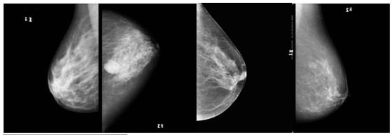
A mammogram is a special, low-dose X-ray technique used to take a picture of the breast, detecting and diagnosing any abnormal lumps or masses in breast tissue. It is one of the best tools for the early identification of breast cancer. With early identification, breast cancer can be cured while in the first stage, and recovery is more likely.

Product Features
1. Adopt specialized mammography flat panel detector digital imaging technology. 2. Full size digital mammography x-ray imaging.3. Unique adopt all-solid-state high frequency high voltage generator.4. The safest mammography at high voltage. There is a built-in X-ray ignition coil in host machine, high-voltage power lines less then 25cm.5. Mammography image acquisition control workstation, DICOM 3.0.6. Electric Isocentric rotating C-arm with a unique automatic back to center function.7. Optional the third generation imported moving grid.8. Optional auto/semi-auto/manual, three kind exposure modes.9. Optional image output device: digital film printer.10. A total of 3 pieces of large size full color LCD screen display, operation table 8 inch LCD screen is a touch key.11. Comfortable Compression: When some degree of pressure is required for radiography, it allows you to presser the appropriate pressure (up to a maximum of 20kg) and is equipped with MICOM Control’s Soft-touch system which is designed to minimize the discomfort of the examine with in the pressure range.Tissue Compression: Manual and Motorized (Max 20kg) / Compression Force and Thickness Data Display / Micro Control’s Compression / Automatic Release
Leave Your Message:
Write your message here and send it to us.


