Data show that the total incidence of birth defects in my country is about 5.6%. Nervous system malformations are one of the most common congenital malformations, with an incidence of about 1%, accounting for about 20% of the total number of congenital fetal malformations.
The structural development of the fetal nervous system determines its neurological function in life after birth. Accurately understanding the developmental law and normal structure of the fetal brain is the basis for diagnosing whether the fetal central nervous system is abnormal or not.
In the past, there was no reference to normal structures, and clinicians often felt isolated and helpless due to a lack of understanding of the normal ultrasound appearance of the fetal brain in a certain cycle, and the lack of reference information such as how structures develop in different cycles. If there is a map of the normal performance of the fetal brain for reference, it will be like a rainy season.
A New Tool for Fetal Brain Ultrasound Diagnosis
“Ultrasonic Anatomy Atlas of Normal Fetal Nervous System Development” is divided into 5 chapters, respectively from the normal embryonic development of the nervous system, the normal ultrasonic anatomy of the nervous system in the middle and late pregnancy, the three-dimensional imaging technology of the fetal brain, and the three-dimensional crystal simulation imaging in the fetal brain. The five aspects of application and fetal nervous system ultrasound measurement and normal reference values elaborately describe the normal fetal nervous system, that is, the normal structure and ultrasound performance of the brain development process, as well as the normal value measurement reference.
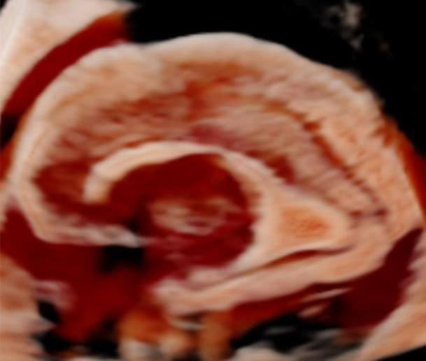
Among them, Samsung’s unique inverted crystal imaging technology has played an important role as a new tool for fetal brain ultrasound diagnosis.
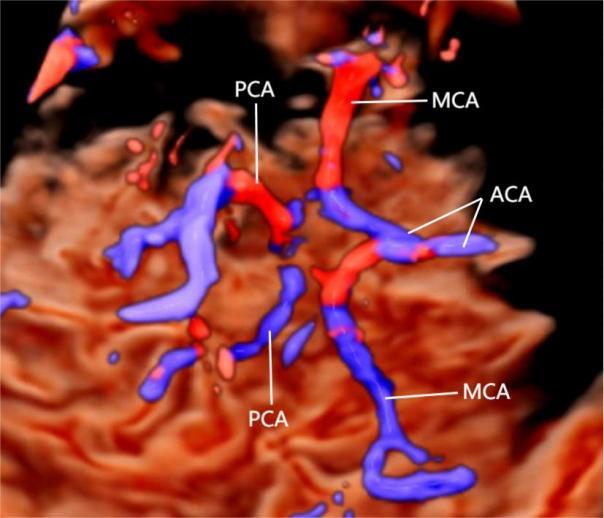
The crystal blood flow imaging mode can superimpose various Doppler color blood flow patterns in the three-dimensional imaging mode to display the position, shape and distribution density of intracranial blood vessels in the tissue. This mode can display the blood flow as a three-dimensional color blood flow image alone or together with the surrounding structures; it provides a new method for the correct evaluation of the fetal cerebral surface sulci and gyrus, and helps doctors make more accurate judgments.
Crystal inversion imaging mode Crystal blood flow imaging mode
Post time: Jul-29-2022


