2.CDFI
·Use of CDFI: Check blood vessels, identify the nature of pipelines,
Identify arteries and veins, show origin and direction of blood flow,
Time phase, reflects the nature of blood flow, indicates fast blood flow speed
Slow, guided spectral Doppler sampling position
1)CDFI regular adjustment content (red text):

2)CDFI infrequently adjusts content
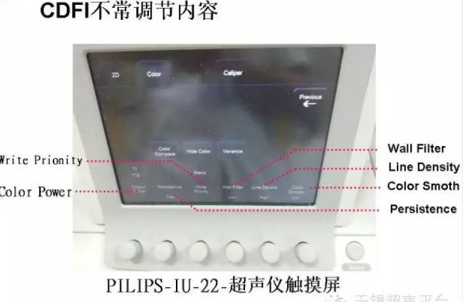
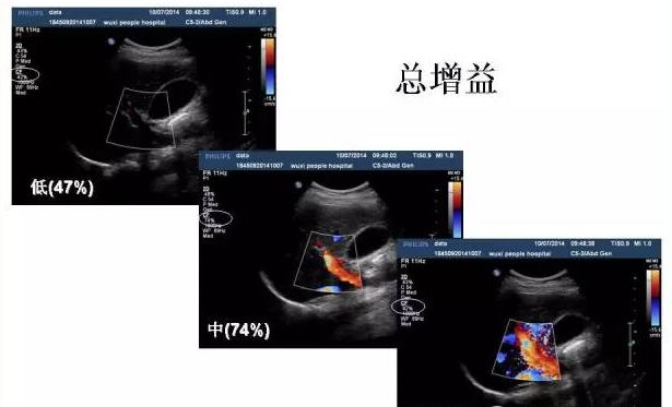
Total gain:
Color box size and position
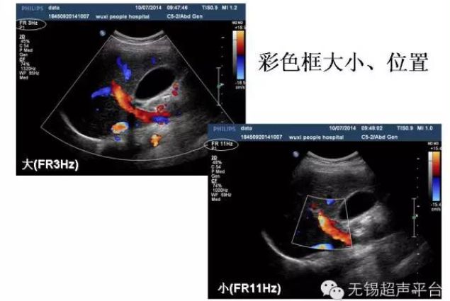
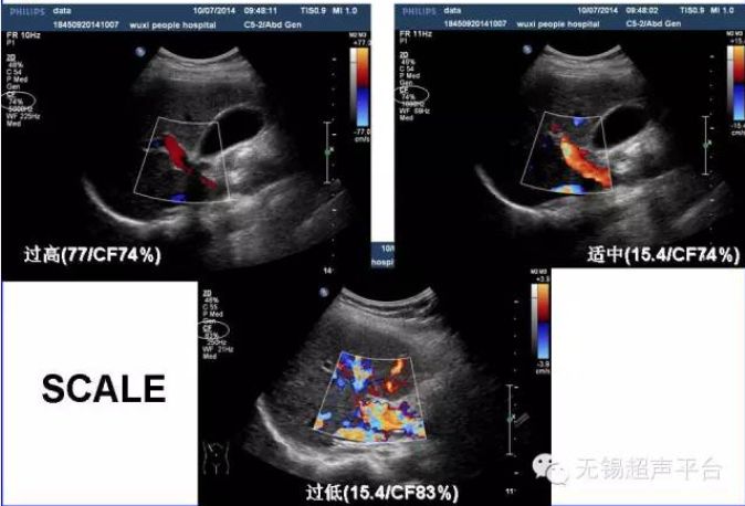
Image difference between too high, too low and moderate Scale
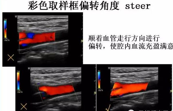
Color sampling frame deflection angle steer
Deflect along the direction of the blood vessel to make the blood flow in the cavity full and satisfactory.
Question 1: How to adjust ultrasound parameters to display low-speed blood flow?
1. Increase---gain
2. Reduce --- speed scale SCALE
3. Add --- sound output Output Power
4. Add --- frame average
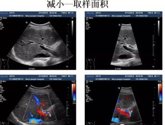
6. Reduce---sampling area
6. Reduce --- the number of focus points (optimize focus)
7. Reduce --- distance
Reduce--sampling area show:
Question 2: How to Reduce color bleeding and remove aliasing?
1. Reduce--gain
2. Add--speed scale SCALE
Question 3: How to increase frame rate?
1. Reduce --- B mode size
2. Reduce --- depth
3. Reduce --- color sampling frame
4. Reduce --- frame average
5. Reduce --- the number of focus points
6. Reduce --- detection distance
3. Spectral Doppler adjustment method
1. Working method: If the flow rate is not high, choose PW, if the flow rate is high, choose CW.
2. Filter conditions: Low-pass filtering is used for low-speed blood flow, and high-pass filtering is used for high-speed blood flow.
3. Speed scale: Select the speed scale corresponding to the detected blood flow speed.
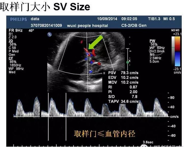
4. Sampling door: detect blood vessels, sampling door ≤ blood vessel inner diameter. Check intracardiac valves
The mouth sampling door is medium sized.
5. Zero baseline: moving the baseline can increase the measurement range in a certain direction and avoid errors.
Now aliased.
6. Frequency shift signal flips up and down: easy to measure, the instrument automatically envelopes the spectrum waveform.
7. Incident angle: cardiovascular examination ≤ 20, peripheral blood vessels ≤ 60, and the angle should be corrected.
8. Transmission frequency: A higher frequency is used for low-speed blood flow, and a lower frequency is used for high-speed blood flow.
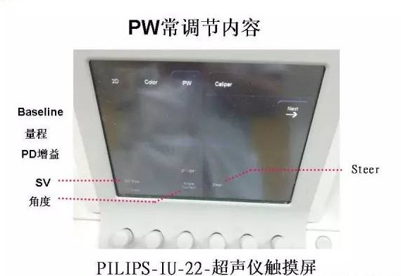
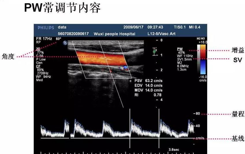
PW often adjusts content
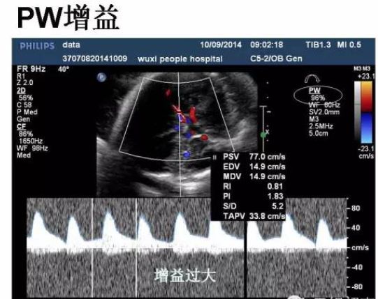
When the PW gain is too large
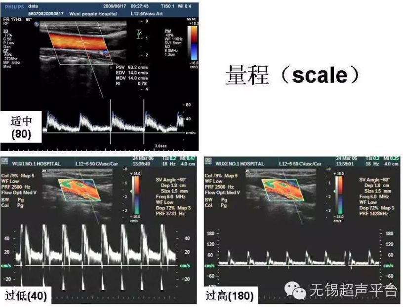
When the range is moderate, too low or too high
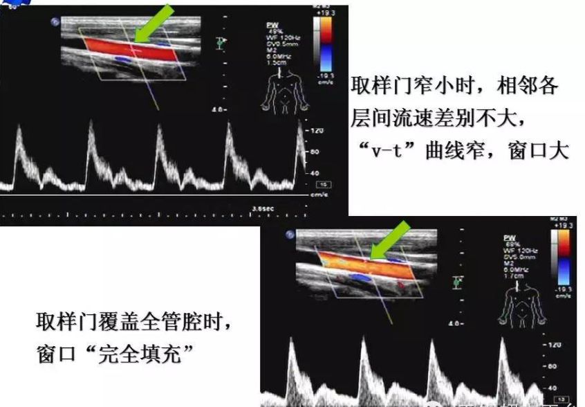
Sampling door size
1. When the sampling door is narrow, there is little difference in flow velocity between adjacent layers, the "v-t" curve is narrow, and the window is large.
2. When the sampling door covers the entire lumen, the window is "completely filled"
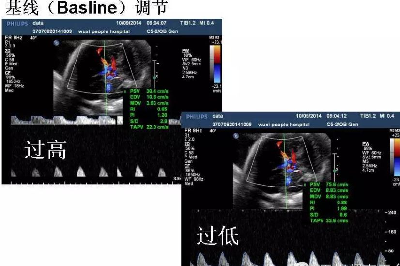
baseline adjustment too high or too low:
Question 5: HoW to Increase PW&CW sensitivity
1. Increase gain
2. Increase sound output
3. Increase sampling volume
4. Set the scanning angle appropriately
Note: Ultrasound instruments have preset conditions and are suitable for examining different organs and tissues.
Based on the preset settings, make appropriate adjustments according to specific conditions to meet diagnostic needs.
Post time: Oct-02-2024


