Home
Products
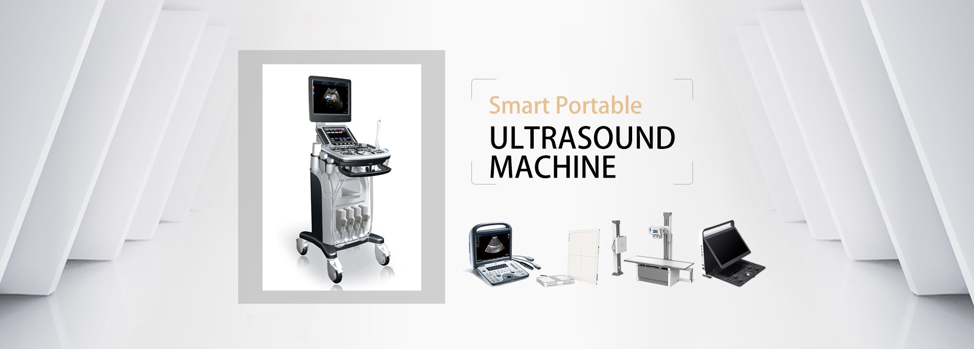
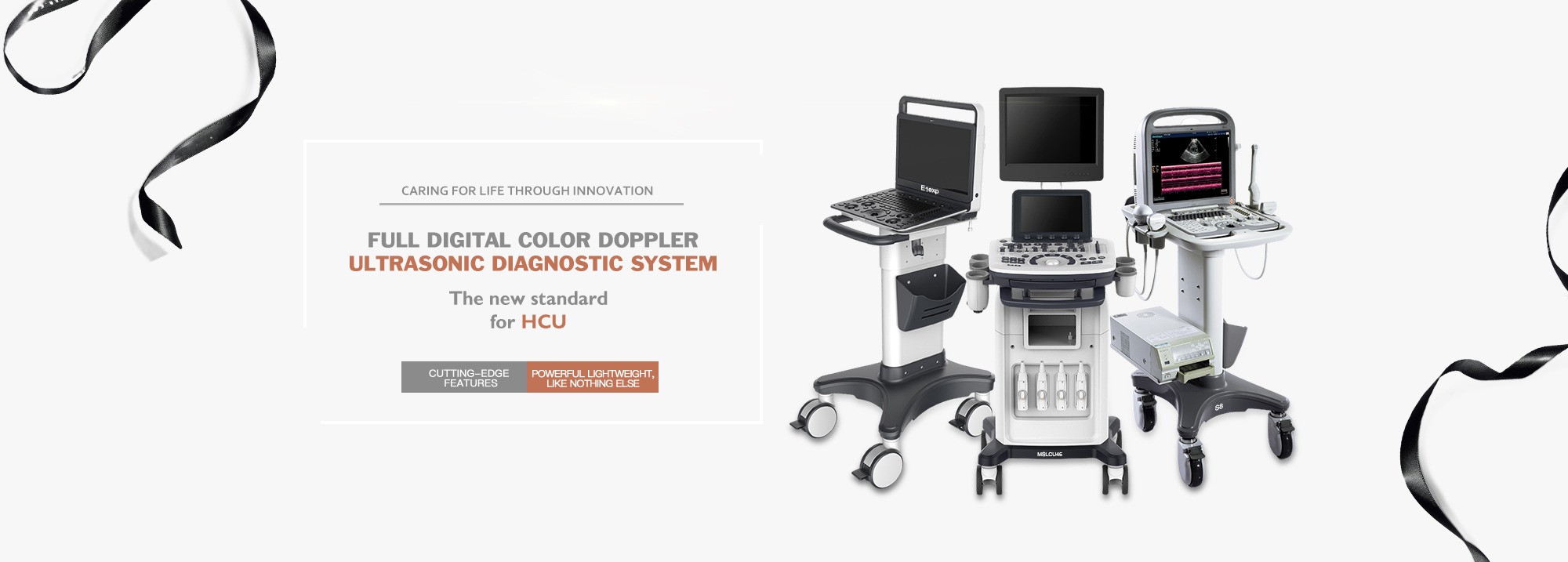
Ultrasound System
Handheld Usb/Wireless Ultrasound
Portable Ultrasound
Cart Ultrasound
Ultrasound Needle/Needle Guide
Sonoscape Ultrasound
Chison Ultrasound
zoncare ultrasound
Mindray Ultrasound
SIUI ultrasound
X-Ray Device
CT
C-arm x-ray machine
DR X-ray machine
Mobile x-ray machine
Portable x-ray
Mammography X-ray system
Radioactivity x-ray system
X-ray film processor
X-ray protection products
OB/GYN, Infant and Gynecology
mammogram machine
Colposcopes
Fetal doppler
Fetal Monitor
Infant Phototherapy
Infant scales
Infant incubator
Infant radiant warmer
Jaundice meter
Gynaecological Bed
Operating Technology and Operating Equipment
Surgery tools, devices
Operating tablets and surgery tablets
Surgery lights, booms
Feeding Pump
Infusion technology equipment
Respiration Equipment, Oxygen Equipment
Sringe pump
Endoscopes and accessories
Anesthesiology equipment, anesthesiology systems
Inhaler and aerosol appliances
Operating Microscope
Suction unit
Medical pendant
Delivery table
Defibrillator
HF (Electrosurgical instruments)
Emergency medicine, first aid, rescue equipment
Emergency medicine first aid
Emergency medicine, first aid, rescue equipment
Hospital Furniture, Equipment, Care Equipment
Hospital Furniture, Equipment, Care Equipment
IVD(In-Vitro Diagnosis), Diagnostic Tests
Mindray Analyzer
Blood Coagulation Analyzer
Clinical Chemistry Equipment
Glycated hemoglobin analyzer
Gynetic testing, Molecular diagnostics
Haematology / histology cytology testing
Immunochemistry testing / immunology testing
Infectious Immunology Testing
Microbiology testing
PCR Analyzer
ENT Equipments
Hygiene / Sterilisation / Disinfection
Autoclaves / Sterilizers
Benchtop Autoclave
Portable Autoclave/Sterilizer
Steam Sterilizer
Packing Machine
Dental Autoclave
Walking and Mobility Aids
Diagnostics Equipments
Blood Glucose Monitors
ECG, Heart Circulation Diagnostics Equipments
Audiomter, Audiological Diagnostics Euipments
Ophthalmic Diagnosis and Ocular Diagnosis
OB/GYN, Gynaecological Equipment, Colposcope
Microscopes, Varioscope
Neurological Diagnosis, Apparatus And Instrument
Sleep Studies Apparatu
Thermometer /Body Temperature Measurement Devices
Urological Investigation And Diagnosi
Scales – Body Fat Testing
Blood Pressure Monitoring Equipments
Blood Flow Measurement Equipments
Vein Finders
Laboratory Equipment
Centrifuges
Hematocrit Centrifuge
High Speed Centrifuge
Refrigerated Centrifuge
Incubators
medical freezer
Microtome
Pipette
See all categories
Dental Equipments
Dental Equipment
Dental Unit
Dental Air Compressor
Dental Ultrasonic Scaler
Dental Chair
Dental Autoclave
Dental sensor
Dental X Ray Machine
Dental Handpiece
Water distiller
Medical sealing machine
Home Care Equipment
Beauty Equipments
Antigen Rapid Test Kit
About Us
FAQs
Founder Story
Solutions
News
Solution
Contact Us
English
Home
News
How to adjust the instrument during ultrasound examination (with step-by-step explanation– Part 1)
How to adjust the instrument during ultrasound examination (with step-by-step explanation– Part 1)
Post time: Nov-23-2023
Phone
Phone
+8617360196190
Email
Email
info@amaintech.com
Whatsapp
Whatsapp
+8617360196190
Send
Send
Send email to us
Leave Your Message:
Write your message here and send it to us.
English
French
German
Portuguese
Spanish
Russian
Japanese
Korean
Arabic
Irish
Greek
Turkish
Italian
Danish
Romanian
Indonesian
Czech
Afrikaans
Swedish
Polish
Basque
Catalan
Esperanto
Hindi
Lao
Albanian
Amharic
Armenian
Azerbaijani
Belarusian
Bengali
Bosnian
Bulgarian
Cebuano
Chichewa
Corsican
Croatian
Dutch
Estonian
Filipino
Finnish
Frisian
Galician
Georgian
Gujarati
Haitian
Hausa
Hawaiian
Hebrew
Hmong
Hungarian
Icelandic
Igbo
Javanese
Kannada
Kazakh
Khmer
Kurdish
Kyrgyz
Latin
Latvian
Lithuanian
Luxembou..
Macedonian
Malagasy
Malay
Malayalam
Maltese
Maori
Marathi
Mongolian
Burmese
Nepali
Norwegian
Pashto
Persian
Punjabi
Serbian
Sesotho
Sinhala
Slovak
Slovenian
Somali
Samoan
Scots Gaelic
Shona
Sindhi
Sundanese
Swahili
Tajik
Tamil
Telugu
Thai
Ukrainian
Urdu
Uzbek
Vietnamese
Welsh
Xhosa
Yiddish
Yoruba
Zulu
Kinyarwanda
Tatar
Oriya
Turkmen
Uyghur